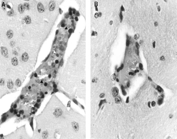
Figure 3.

Histology of the brain (original magnification, 20 × 1.6) shows accumulation of parasitized erythrocytes (open arrow) and infiltration of lymphocytes (dark arrow) within cerebral vessels of BALB/c (A) but not in DBA/2 (B) mice after infection with P. yoelii (17XL strain).
