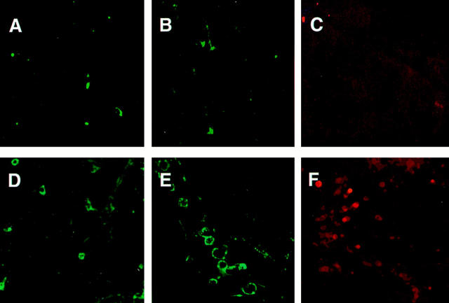
Figure 9.
Confocal microscopy performed on vibratome sections of the brain from mice that received IL-2 treatment and were infected with P. yoelii (strain 17XL) for 6 days. Sections of brain from a mouse given IL-2 treatment but no infection, and stained with anti-αβ TCR antibody (A), with anti-γδ TCR antibody (B), or with anti-CD54 (ICAM-1) antibody (C); no marked staining was seen in any of the cases. Sections of brain from a mouse given IL-2 treatment and infection, and incubated with anti-αβ TCR antibody (D), with anti-γδ TCR antibody (E), or with anti-CD54 antibody (F); a number of cells with positive staining was seen in each cases, notably with anti-γδ TCR antibody. Laser power, PMT gains, and confocal thresholds were set using FITC-IgG isotype controls and kept constant throughout the experiment.

