Figure 1.

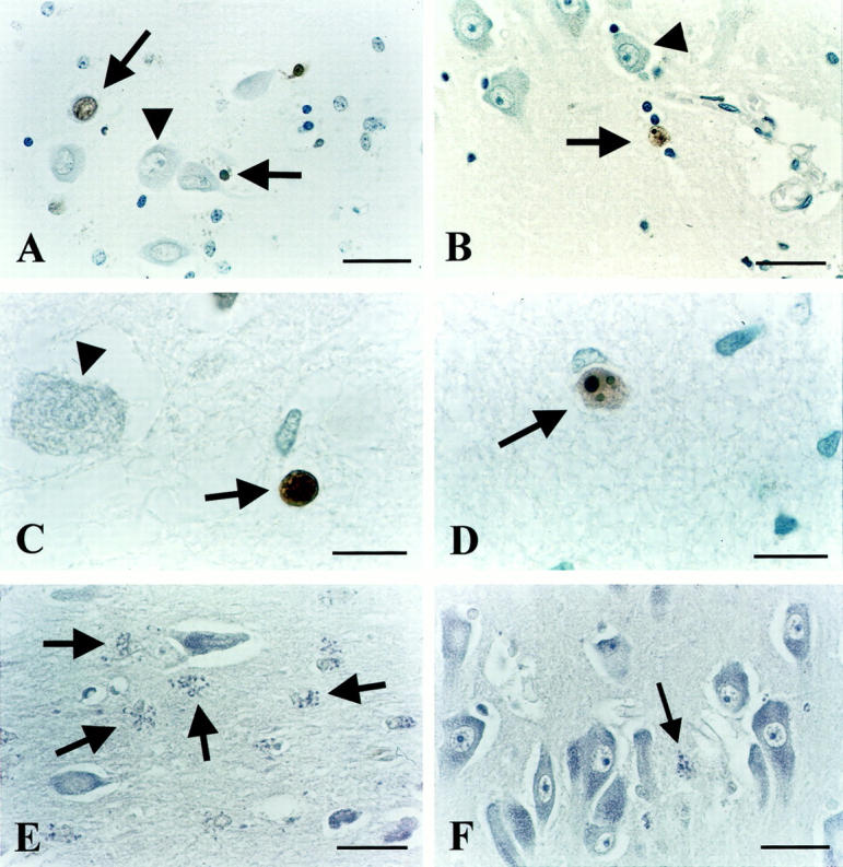
ISEL results. A: Positive labeling in the CA4 area of depressed patient 94-112, showing necrotic morphology (upper arrow) as indicated by the comparable size as an intact, neighboring neuron (arrowhead) without chromatin re-organization or apoptotic bodies visible. Also seen is a labeled apoptotic cell as evidenced by its pycnotic appearance, strong condensation, and brown DAB precipitate (horizontal arrow). B: ISEL-positive neuron (arrow) just outside the CA1 cell layer of depressed patient 90-001 with clear apoptotic morphology, ie, a reduced size as compared to unstained, healthy-looking neurons (triangle), and apoptotic bodies clearly visible. C: ISEL-positive, apoptotic cell (arrow) with a pycnotic, condensed appearance adjacent to a nonstained large cell (arrowhead). CA1 of depressed patient 94-094. D: Apoptotic neuron (arrow) in the subiculum of depressed patient 94-032 with three clear apoptotic bodies visible. E: Frequent, granular morphology (arrows) suggestive of chromatolytic processes, adjacent to normal looking neurons in CA3 of depressed patient 90-001. F: Normal-appearing neurons in CA1 of control subject 94-123. Also, one granular, chromatolytic-like structure is visible (arrow). Scale bars: 34 μm (A, B, E, and F) and 15 μm (C and D).
