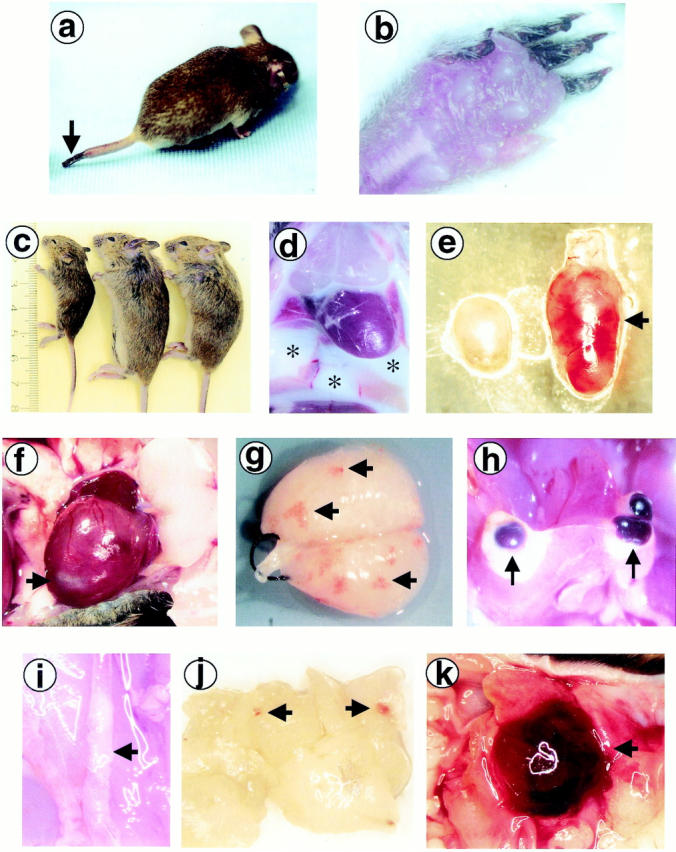
Figure 1.

Gross appearance of PC−/−/FXI−/− animals. a and b: I107, the oldest surviving PC−/−/FXI−/− mouse (94 days), presented with gangrene on the tail tip (a; arrow) and toes (b), and severe inflammation and edema on the paws (b). c: The majority of the PC−/−/FXI−/− animals (left) were significantly growth retarded compared to their PC+/−/FXI−/− (middle) and PC+/+/FXI−/− (right) littermates. d: G352-2 and G506, two of the four PC−/−/FXI−/− animals discovered dead, presented with a significant volume of milky-white lymphatic fluid in the thoracic cavity (*). The population of cells was primarily lymphocytic. e: A common attribute observed in these animals was enlargement and apparent hemorrhage of lymph nodes (arrow). f: Hearts were also affected with various anomalies including infarcted tissue (arrow). g: After perfusion, lungs generally demonstrated hemorrhagic lesions (arrows). h: Hemorrhagic cysts were commonly found adjacent to fibrotic areas in the livers (arrows). i: Atherosclerotic areas were seen in some of the larger vessels such as the abdominal and thoracic aorta (arrow). j and k: Soft tissue hemorrhage (arrows) was also commonly observed, as seen in the pancreas (j) and the preputial gland (k).
