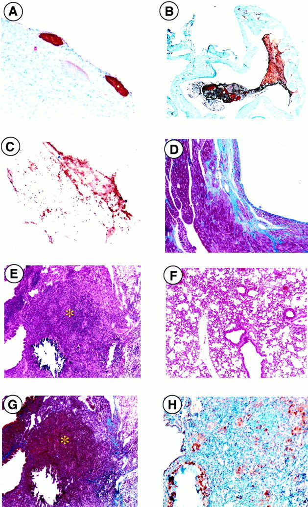
Figure 2.

Microscopic analysis of PC−/−/FXI−/− hearts and lungs. Thrombus formation was readily observed (brown areas) via antifibrin(ogen) immunostaining in various regions of the heart, such as the epicardium (A) and atria (B). C: Anti-P-selectin immunostaining identified the presence of activated platelets within the thrombus. D: Ventricular fibrosis, seen macroscopically in Figure 1F ▶ , was verified through Masson’s Trichrome staining in blue. E: Lung tissue demonstrated focal areas of consolidation and mineralization (*) (H&E) as compared to a PC+/+/FXI−/− (F) control. G: Collagen deposition (blue) observed by Masson’s trichrome staining. H: Macrophage infiltration (brown) visualized by anti-Mac3 immunostaining was also observed. Original magnifications: ×40 (B), ×100 (A, D–F, G), ×200 (H), and ×400 (C).
