
Figure 1.
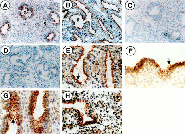
Representative illustration of IL-1RII immunostaining in the human endometrium. Sections of endometrial tissue were incubated with mouse monoclonal anti-IL-1RII antibody (A, proliferative day 13; B, secretory day 24; original magnification, ×68) or with an equivalent concentration of normal mouse IgGs (C and D, respectively; original magnification, ×68). Sections were then incubated successively with biotinylated goat anti-mouse polyclonal antibody and avidin-biotinylated horseradish peroxidase complex. The immunoreaction was revealed with diaminobenzidine (brown staining) and hematoxylin was used for counterstaining (blue staining). Note the brown fine positive staining in stromal and epithelial cells (cellular staining) (E–H; original magnification, ×268), and the brown deposit (arrow) that is primarily located at the apical side of glandular (E, secretory phase day 24) and surface (F, secretory phase day 16) epithelium, or more spread within the glands lumen (G, secretory phase day 16). Positive immunostaining is also detected in isolated stromal cells (c) (G, secretory phase day 16) and microvessels (v) (H, secretory phase day 24) found in the stroma in the secretory phase of the menstrual cycle. s = stroma, g = gland.