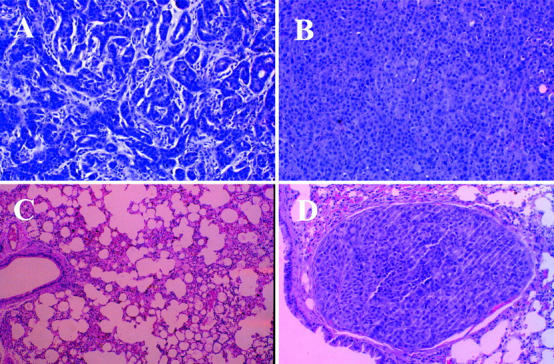
Figure 4.

Histology of sections taken from mouse primary tumors and lung tissues. Sections taken from primary tumors and lungs of female athymic nu/nu mice orthotopically injected with either MCF-7 or MDA-435 cells were fixed in formaldehyde and embedded in paraffin. The slides were stained with H&E and visualized at ×40. A: MCF-7 adenocarcinoma, site of inoculation. B: MDA-435 adenocarcinoma, site of inoculation. C: MCF-7 lung, absence of metastases. D: MDA-435 lung, lumen of blood vessel is occluded by tumor embolus.
