
Figure 2.
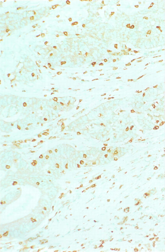
Immunohistochemistry for CD3 pan-T cell marker with methyl green counterstain. Numerous T lymphocytes are present in the neoplastic epithelium of this MSI-H colon carcinoma as well as in the surrounding nonneoplastic stroma. (Same specimen as Figure 1 ▶ .)