
Figure 11.
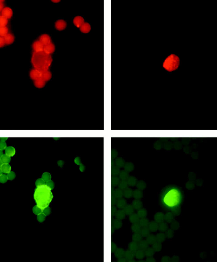
Representative dual-labeling studies of CECs stained with the antibody for HO-1 (red staining, top left and right) and with the endothelium-specific antibody, mAb-P1H12, (green staining, bottom left and right). Top left: Faint red staining with HO-1 antibody in an endothelial cell from a control subject, and designated “low positive” in scoring summarized in Figure 12 ▶ . Top right: Strong red staining with HO-1 antibody in an endothelial cell from a sickle patient, and designated “high positive” in scoring summarized in Figure 12 ▶ . Bottom left: Green staining with mAb-P1H12 of the cell shown in top left demonstrating that this is an endothelial cell. Bottom right: Green staining with mAb-P1H12 of the cell shown in top right demonstrating that this is an endothelial cell.