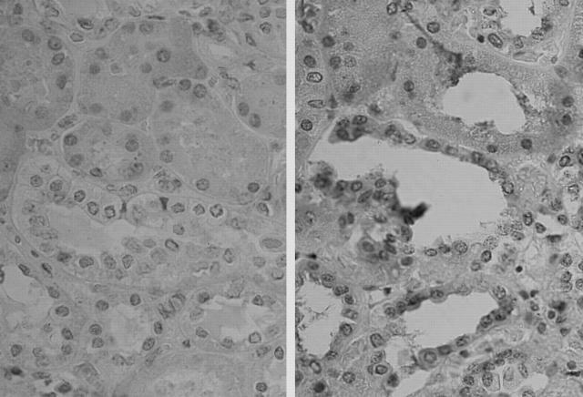
Figure 9.
Immunoperoxidase staining for HO-1 in the kidney of a patient with sickle cell disease. Immunoperoxidase studies were undertaken in the absence (left) and presence (right) of HO-1 antibody. Right: Positive staining for renal distal tubules and interstitial cells in the sickle kidney. Original magnification, ×400.

