Figure 1.

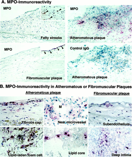
Expression of MPO immunoreactivity in various stages of human atherosclerotic arteries and localization in advanced plaques. Frozen sections were incubated with pAb-MPO and MPO immunoreactivity was visualized with the alkaline-phosphatase ABC method (red). A: MPO immunoreactivity localized in early to advanced human atherosclerosis (arrowheads). Atheromatous plaques exhibited abundant MPO, but lacked staining by nonimmune rabbit IgG used as a negative control. Arteries with fatty streaks, and fibromuscular plaques usually exhibited little MPO (large panels), but some samples contained MPO in the intima (insets). Original magnification, ×200. These results are representative of 14 fatty streaks, 17 fibromuscular plaques, and 25 atheromatous plaques. B: MPO-containing cells were localized in all regions of advanced plaques, especially in fibrous cap, near microvessel (M, indicates lumen of microvessel), and lipid core of atheromatous plaques. Some of fibromuscular plaques contained MPO-containing cells in the subendothelial space and deep intima. Some of the lipid-laden foam cells were also MPO-positive in atheroma (arrowheads). Original magnification, ×400.
