
Figure 4.
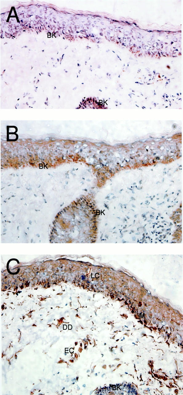
Transmembrane fractalkine is predominantly expressed in the epidermis of noninflamed human skin. Noninvolved skin samples were taken after resection of human facial skin tumors. Frozen sections (8 to 10 μm) were cut for analysis by immunohistochemistry. Immunohistochemical labeling was performed using anti-fractalkine and anti-peptide polyclonal reagents. A: The staining using the goat α-Fkn revealed positive cells restricted to the basal keratinocytes of the human epidermis, with no obvious positive cells within the dermis. B: Staining using rabbit α-C pep, which reacts to an intracellular epitope of fractalkine, revealed a similarly restricted staining pattern, again with no obvious positive cells within the dermis. C: Staining using the rabbit α-N-pep reagent showed discrete positive cell staining within the epidermis with morphology characteristic of Langerhans cells (a single cell is emphasized against the background), along the basement membrane of the epithelium with characteristics of melanocytes (M), there was also discrete staining of cells within the dermis, including structures with the appearance of blood vessels (EC) and dermal dendrocytes (DD). Negative control reagents showed no background staining (data not shown). Original magnifications, ×400.