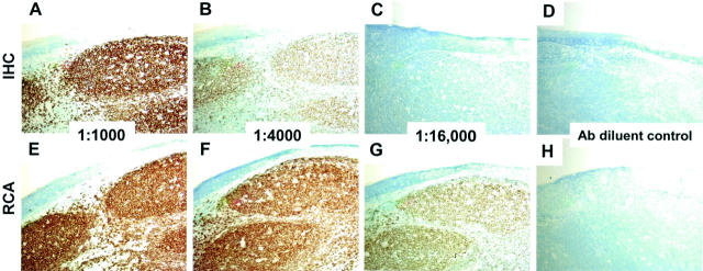
Figure 2.
Detection of CD20 by conventional immunohistochemistry or immuno-RCA. Serial sections of formalin-fixed paraffin-embedded tonsil were prepared for immunohistochemistry as described in the text and incubated with monoclonal antibody to CD20 diluted 1:1000 (A and E); 1:4000 (B and F); 1:16,000 (C and G); or antibody diluent alone (D and H). After development with a standard detection kit (A–D) or by immuno-RCA (E–H) using an RCA conjugate that recognizes mouse immunoglobulins, sections were incubated with diaminobenzidine/hydrogen peroxide (brown stain) and counterstained with methylene blue. All panels, showing the same region of the tonsil specimen, were photographed at an original magnification of ×10. Uniform staining was observed in the germinal centers, whereas the interfollicular regions of the tonsil showed little staining. IHC, immunohistochemistry.

