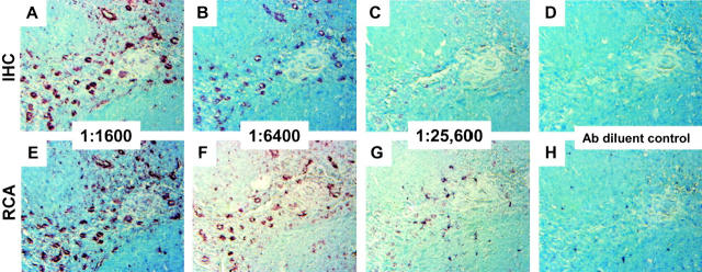
Figure 4.
Detection of vimentin. Serial sections of formalin-fixed paraffin-embedded tonsil were prepared for immunohistochemistry as described in the text and incubated with monoclonal antibody to vimentin diluted 1:1600 (A and E); 1:6400 (B and F); 1:25,600 (C and G); or antibody diluent alone (D and H). After development with standard immunohistochemistry techniques (A–D) or immuno-RCA (E–H), sections were incubated with Vector Nova Red/hydrogen peroxide (brown stain) and counterstained with methylene blue. All panels, showing the same region of the tonsil specimen, were photographed at an original magnification of ×20. In contrast to CD20 (Figure 2) ▶ , lymphoid and endothelial cells in the interfollicular areas stain positively for vimentin. IHC, immunohistochemistry.

