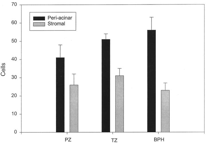
Figure 2.
Quantitation of the localization of FGF7-expressing cells in prostate tissue. Frozen sections of six normal peripheral zone, six normal transition zone, and six BPH tissues were analyzed by immunohistochemistry with anti-FGF2 monoclonal antibody. The number of cells located adjacent to epithelial acini (periacinar) was compared to the number of cells in stromal areas using a point-counting protocol. The mean counts in the 10 fields (± SEM) in each location for normal peripheral zone (PZ), normal transition zone, and hyperplastic transition zone (BPH) are shown. For all three types of tissue the difference between the periacinar and stromal tissues is statistically significant (P < 0.04, paired t-test).

