
Figure 1.
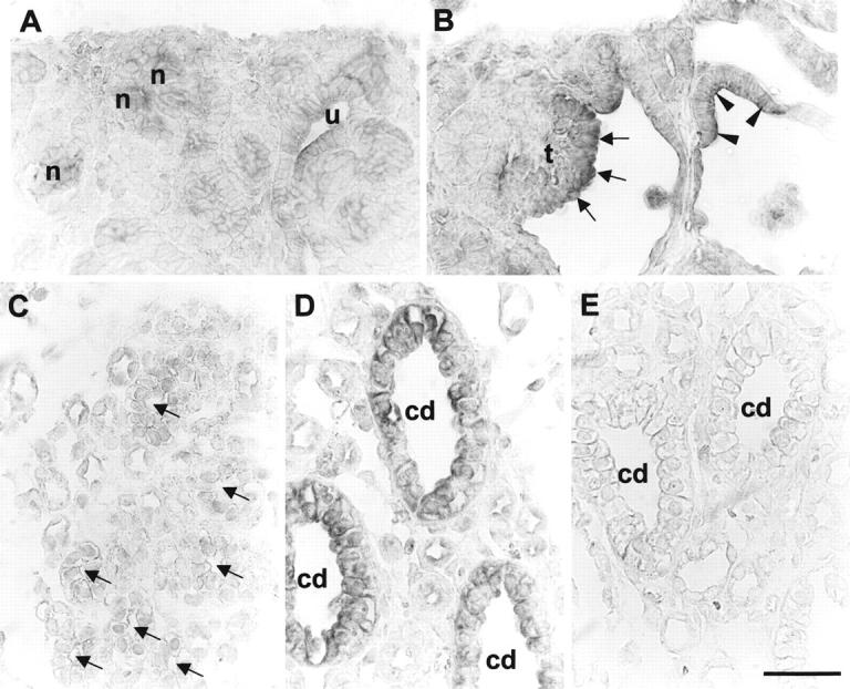
TGF-β 1 in situ hybridization in sham-operated and obstructed ovine fetal kidneys. A and C are sections of sham-operated developing kidneys, whereas B, D, and E are from obstructed kidneys. All panels represent the results of hybridization with TGF-β1 antisense probes, apart from E in which a control sense probe was used. A: TGF-β1 transcripts were detected in ureteric bud (u) and forming nephrons (n) in the normal superficial cortex. B: Prominent signal for TGF-β1 was observed in dilated tubule epithelia (arrowheads) and glomerular tufts (t; arrows) in the obstructed cortex. C: Faint TGF-β1 signal was detected in sham-operated developing medullary collecting ducts (arrows). D: In contrast, there was TGF-β1 transcript up-regulation in the larger dilated collecting ducts (cd) in the obstructed kidneys. E: Only background signal was detected using the sense probe. Scale bar, 15 μm.