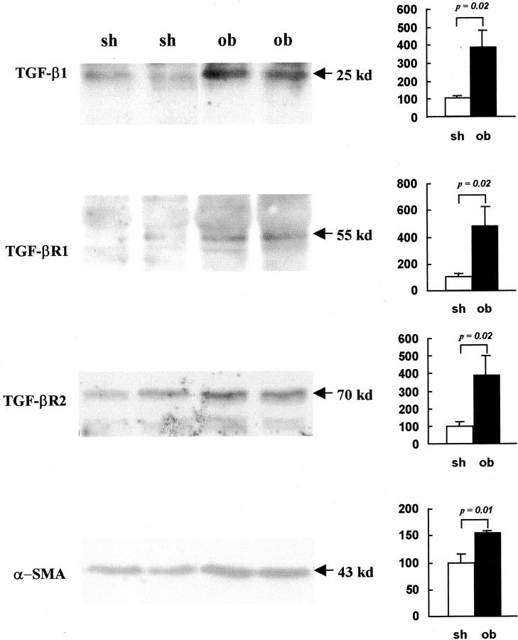
Figure 5.
Western blot of TGF-β1, its receptors and α-SMA protein in sham-operated and obstructed ovine fetal kidneys. Representative Western blots showing protein extracts from two sham-operated (sh) and two obstructed (ob) kidneys probed with antibodies to TGF-β1, TGF-βR1, TGF-βR2, and α-SMA. Bands at the expected sizes were detected in each Western blot (left). Signal intensity, measured by densitometry, was compared in sham-operated and obstructed samples (n = 5) using a Student’s t-test (right). This analysis demonstrated that obstruction was associated with significant up-regulation of protein expression as follows: TGF-β1 (281% greater than control, P = 0.02), TGF-βR1 (379% greater than control, P = 0.02), TGF-βR2 (291% greater than control, P = 0.02), and α-SMA (54% greater than control, P = 0.01).

