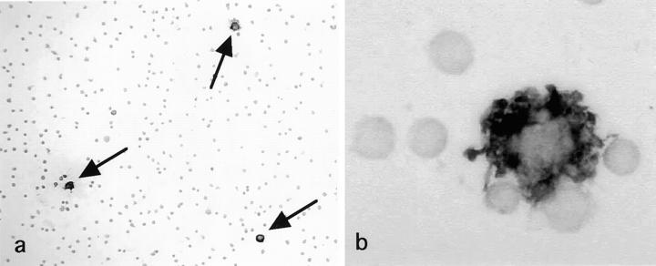
Figure 2.
S-100b-immunoperoxidase staining applied to fresh smeared mesenteric LN cells. S-100b protein is detected specifically in the cytoplasm of IDC. a: Approximately 0.6% cells corresponded to S-100b+ IDC, which were larger than lymphocytes and tend to attach several lymphocytes. Arrows indicate S-100b+ IDC. Magnification, ×100 b: A high power view of S-100b+ IDC. Note intense S-100b+ immunoreaction products in the cytoplasm of IDC attaching to a few lymphocytes. Magnification, ×1700.

