
Figure 1.
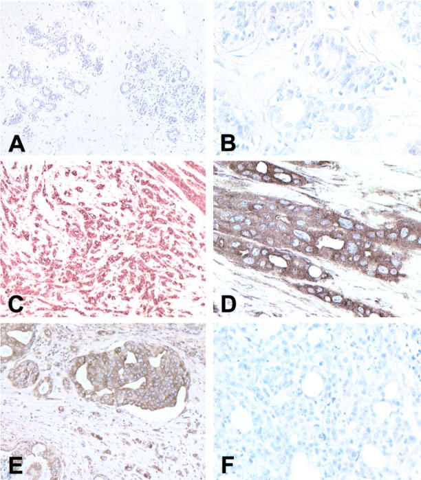
MT-3 staining in the human breast. MT-3 antibody was used at a 1:200 dilution and all slides were counterstained with hematoxylin. A: Normal breast epithelium, both ducts and lobules, does not stain for MT-3. Original magnification, ×100. B: Higher power of normal breast ducts reveals absence of staining for MT-3. Original magnification, ×400. C: Case of invasive duct carcinoma which stains strongly for MT-3. Original magnification, ×100. D: Higher power of that in C demonstrating the strong cytoplasmic MT-3 staining. Original magnification, ×400. E: Area of DCIS and adjacent invasive duct carcinoma, which stains strongly for MT-3. Original magnification, ×200. F: Case of invasive ductal carcinoma that does not stain for MT-3. Original magnification, ×200