
Figure 2.
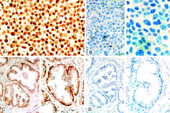
Competitive ER-β immunostaining DU145 cells (A and B) and normal prostate (C and D). A: DU145 cells immunostained in the absence of competing peptide are shown. Note the strong nuclear staining in the majority of cells (original magnification, ×265). B: After incubation of GC-17 with 40 μg of the immunizing peptide, there was almost a total absence of cells with positively stained nuclei (compare with A) [original magnifications: ×115 (left), ×350 (right)]. C: Tissue section of normal prostate. In the absence of competing peptide strong immunostaining of cells in the basal layer of glands is seen in this section of prostate (see also Figure 4, A and B ▶ ) [original magnifications: ×115 (left), ×230 (right)]. D: A replicate section of the prostate illustrated in C after preincubation of GC-17 with 40 μg of immunizing peptide. Note the absence of immunostaining in these sections [original magnifications: ×90 (left), ×280 (right)]. For details of the procedures used in these studies see Materials and Methods. All sections were counterstained with 10% Harris hematoxylin.