
Figure 6.
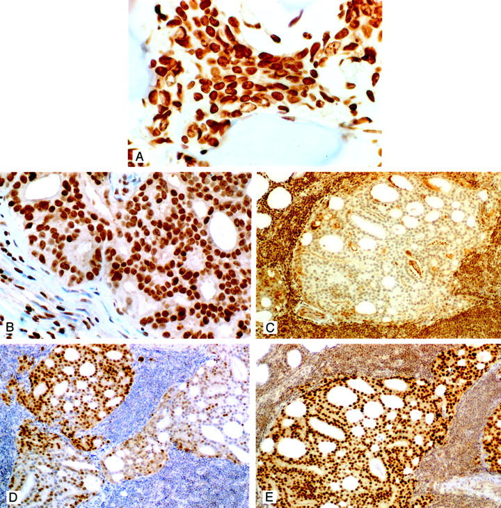
A: ER-β immunostaining in a prostatic carcinoma metastatic to bone. Note the strong nuclear immunostaining for the β receptor in this metastatic lesion. The neoplastic cells are localized between spicules of bone. Strong to moderate staining was present in the majority of metastases to bone (original magnification, ×400). B: ER-β immunostaining in a prostatic carcinoma metastatic to an internal iliac lymph node. Strong nuclear immunostaining is evident in this metastatic lesion. Strong PSA immunostaining of these cells were found (not illustrated). Note that the nuclei of several stromal cells in this lymph node are also positively stained for the receptor (original magnification, ×400). C: ER-β immunostaining of a prostatic carcinoma metastatic to a lymph node. In this example the metastatic cells were unstained for the receptor. Cells in this same cancer were however immunostained for ER-α and AR (see D and E). As was the case for the metastasis illustrated in B these cells were strongly PSA-positive. Note that lymphocytes are strongly stained for the receptor, which was a consistent finding and provided an internal positive control for immunostaining with the β antibody (original magnification, ×100). D: ER-α immunostaining of the same lesion illustrated in C. Immunopositive cells are seen scattered throughout this metastatic lesion. In one other case of lymph node metastasis a very few positive cells (<10%) were present. ER-β was however strongly expressed in that metastatic lesion. Note the absence of ER-α immunostaining of lymphocytes, a consistent finding that was in contrast to ER-β staining in these cells (original magnification, ×100). E: Representative section of AR immunostaining in metastatic lymph node lesions. Strong nuclear staining was uniformly found in the majority of cells that comprised these metastatic lesions. Cytoplasmic staining occurred only in metastases from the patient who had received anti-androgenic therapy. Lymphocytes were lightly stained for AR (original magnification, ×100). All sections were counterstained with 10% Harris hematoxylin.