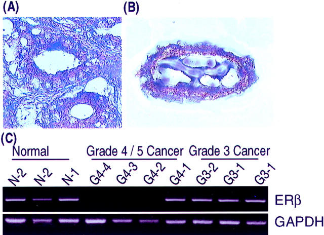
Figure 7.
A and B: LCM dissection of a grade 3 carcinoma. A: The grade 3 neoplastic gland that was microdissected is seen in the center of this microscopic field (original magnification, ×325). B: The microdissected lesion is seen on the transfer cap (original magnification, ×325). Both sections were lightly stained with 10% Harris hematoxylin. C: Results of RT-PCR analysis of ER-β mRNA on microdissected human normal prostate acini (N-1 and N-2) and grade 3 (G3–1 and G3–2) and 4/5 (G4–1 to G4–4) carcinomas. Total RNAs were extracted from the microdissected tissues on transfer cap and reverse-transcribed. The resultant cDNAs were subjected to PCR analyses. The amplified products were run into 2% agarose gel with ethidium bromide. Representative fluorographs for ER-β and GAPDH RT-PCR analyses are shown.

