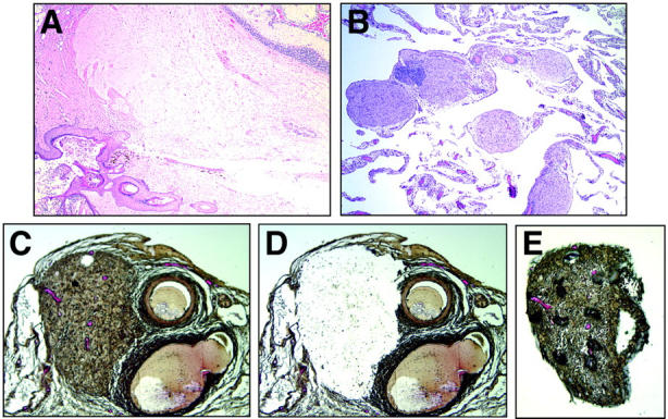
Figure 1.

A: H&E-stained section of a mature teratoma containing keratinizing squamous epithelium and hair follicle (bottom left), mature glial tissue (center), and cerebellar tissue (top right) (original magnification, ×40). B: H&E-stained tissue section of omentum. Five individual foci of mature glial tissue are pink and round to ovoid in shape (original magnification, ×100). C: Modified H&E-stained section showing a glial implant (left) and adjacent blood vessels (right) before laser-capture microdissection (original magnification, ×200). D: Same section as shown in C after laser-capture microdissection (original magnification, ×200). E: Captured glial implant. The dark bubbles overlying the implant are an artifact of the capture membrane (original magnification, ×200).
