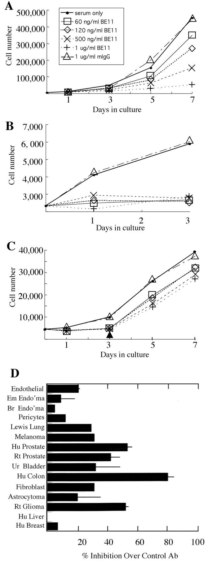
Figure 4.

Anti-βE11 inhibits tumor cell growth in vitro. Primary and tumor cells were grown as described in Materials and Methods in the presence or absence of anti-βE11 or a murine IgG control for the number of days indicated. A: Cx.1 human colon carcinoma cells. B: LNCaP human prostate tumor cells. C: LNCaP cells were grown as in B for 3 days after which antibody was washed out of the medium (indicated by arrowhead). Legend in B and C is the same as that for A. D summarizes the maximal inhibition observed for each cell line used in the growth assays. For all cell lines, maximal inhibition occurred at days 5 to 7 and at a βE11 antibody concentration of 0.5 to 1.0 μg/ml. From top to bottom, cell lines are as follows: primary bovine retinal endothelial cells, murine embryonic endothelioma, murine brain endothelioma, primary bovine retinal pericytes, Lewis Lung carcinoma, B16 BL6 murine melanoma, LNCaP human prostate, rat prostate, human urinary bladder carcinoma, Cx.1 human colon carcinoma, murine 3T3 fibroblast, human astrocytoma, C6 rat glioma, HEPG2 human liver carcinoma, and MCSF7 human breast carcinoma.
