Figure 5.
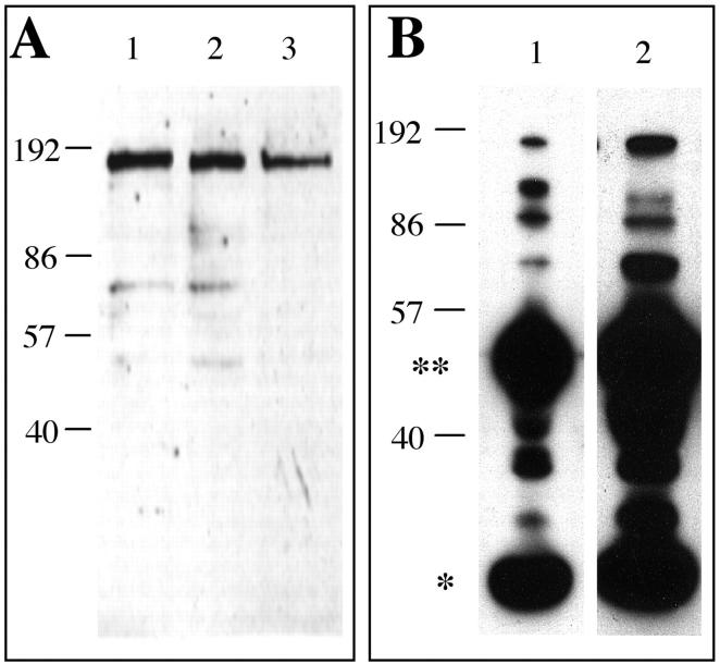
The βE11 antigen is expressed in serum-free melanoma cells. A: Western blot of detergent lysates (∼10 μg total protein) from LNCaP cells grown in serum (lane 1) and B16 BL6 melanoma cells grown in the presence (lane 2) or absence (lane 3) of serum blotted with anti-βE11 followed by goat anti-mouse IgG-HRP. B: Immunoprecipitation from LNCaP cells grown in serum (lane 1) or B16-BL6 melanoma cells grown in the absence of serum (lane 2) was performed with serum-free affinity-isolated anti-βE11 and goat anti-mouse IgG(Fc)/Sepharose as described in Materials and Methods. The immunoprecipitated complexes were run on 8.5% SDS-PAGE and transferred to nitrocellulose. The blots were probed with anti-βE11 followed by goat anti-mouse IgG-HRP. Double asterisk indicates IgG heavy chain; single asterisk indicates IgG light chain. Molecular weight marker (kilodaltons) appear along left hand of each panel A, B.

