
Figure 1.
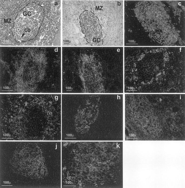
Morphology of intrathyroidal secondary LFs in AITD. a: A section from a formalin-fixed paraffin-embedded block from a HT gland. b to h: Sections from frozen blocks from GD glands. g was deconvolved to improve image definition (see text for details), the other images are standard micrographs. a: H&E staining showing a typical secondary follicle with a GC and a well-formed MZ (cb, centroblasts undergoing mitosis). b: CD20 on B cells of the GC (brown) using the immunoperoxidase technique and counterstained with hematoxylin (GD, case TB228). c: Direct immunofluorescence with peanut agglutinin-fluorescein isothiocyanate, showing positive staining of centroblasts and follicular dendritic cells (GD, case TB378). d: Staining for CD3+ reveals abundant T cells in the MZ with scattered cells inside the GC (GD, case TB228). e: Demonstration of abundant CD4+ T among the T cells in the MZ (GD, case TB228). f: Staining for CD8 shows moderately abundant CD8+ T lymphocytes in the MZ (GD, case TB378). g: Staining for CD 83 highlights the network of mature dendritic cells in the MZ (GD, case TB278). h: Staining for the long form of CD21 reveals the network of follicular dendritic cells in the CG and their polarization toward the light zone (GD, case TB373). i: CD38, as marker of centrocytes. j: CD23 staining revealing the area occupied by the GC. k: CD77 staining as an additional GC marker .