
Figure 3.
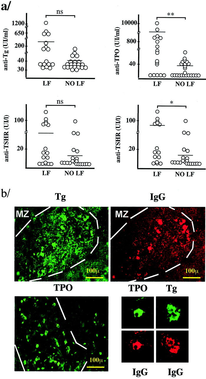
a: Relationship between thyroid antibody titer and the presence of intrathyroidal LFs. TSHR, thyrotropin receptor; LF, group of thyroid glands containing LFs; NO LF, group of thyroid glands without LFs. The plotted data, except for those at bottom right, correspond to all of the patients in Table 2 ▶ . At the bottom right, only the data from GD patients were plotted (*, P < 0.05; **, P < 0.01; ns, not significant P value, Mann-Whitney test). b: Demonstration of the specificity of B and plasma cells in the intrathyroidal LFs. Double immunofluorescence using biotinylated Tg (green, top left) and anti-IgG (red, top right), demonstrating binding of Tg to the same cells that are stained for IgG. Positive lymphocytes only appear in the LFs, whereas plasma cells are present both in the LFs and in the diffuse infiltrate. Bottom left: Binding of biotinylated TPO to abundant cells in the LFs and also to some cells in the diffuse infiltrate. Note the presence of membrane and cytoplasmic staining that correspond to lymphocytes and plasma cells, respectively. Bottom right: Examples of plasma cells stained for TPO and Tg in double immunofluorescence with IgG (GD, case 378).