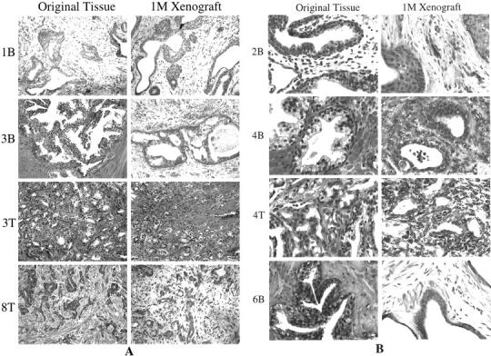
Figure 1.

Histological features of original tissues and corresponding 1-month xenograft tissues are similar, as shown by H&E analysis of representative cases. A: Low-power images (original magnifications, ×1000) were composed so that overall tissue architecture may be evaluated. B: Composed of higher power images (original magnifications, ×2000) for observation of cellular features. Although the majority of glands within each xenograft recapitulated the original tissue, some xenografts contained isolated glands characterized by transitional cell metaplasia (1B) or squamous cell metaplasia (2B, 2T, 3B). Basal cell hyperplasia was also a common feature of the benign xenografted tissues.
