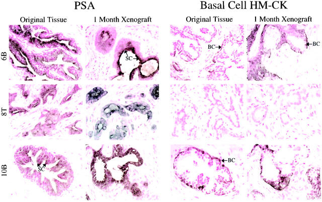
Figure 4.
Expression of PSA and basal cell-specific cytokeratins (HM-CK) were evaluated in the original tissues and matched 1-month xenograft tissues. Representative samples are shown. Benign prostate tissues and xenografts (6B, 10B) were characterized by expression of PSA by the secretory epithelial cells (SC) (dark-brown to black cytoplasmic staining), and distinct expression of HM-CK by the basal cells (BC) lining the glandular structures (brown cytoplasmic staining). Original CaP tissues and corresponding xenografts (8T) were characterized by strong PSA expression by epithelial cells, and a lack of HM-CK (+) basal cells.

