
Figure 2.
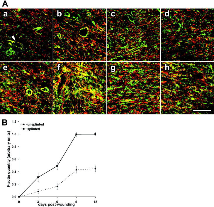
Mechanical tension enhances expression of F-actin and ED-A FN in wound granulation tissue. A: Sections of 3-day-old (Aa, Ae), 6-day-old (Ab, Af), 9-day-old (Ac, Ag), and 12-day-old (Ad, Ah) granulation tissue were double-stained for F-actin (green) and ED-A FN (red) and examined by confocal laser-scanning microscopy. Tissues from unsplinted wounds (Aa–Ad) are compared to those from splinted wounds (Ae–Ah). B: F-actin expression in unsplinted (dashed line) and splinted granulation tissue (continuous line) from 3 days to 12 days assessed by image analysis of immunostained tissue sections (normal dermis = 0 days). In 3-day-old unsplinted granulation tissue (Aa) fibroblastic cells exhibit low levels of de novo-appearing F-actin and ED-A FN whereas fibroblasts in 3-day splinted tissue (Ae) already show important expression and co-localization of the two proteins (yellow); co-localization is also seen in vascular smooth muscle cells (Aa, arrowhead). Expression of both proteins gradually increases and with partial co-localization at 6 days to 12 days after wounding. At any wound age fibroblastic cells of splinted wound granulation tissue exhibit higher expression of ED-A FN and F-actin compared to control (B). Scale bar, 50 μm.