
Figure 3.
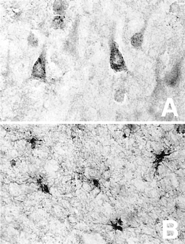
Cystatin C is present in both neurons and glia of AD brain. Temporal cortex of AD brain immunostained for cystatin C revealed a vesicular pattern in pyramidal neurons (A) as well as staining in activated glia in the white matter (B). Original magnifications, ×500.