
Figure 5.
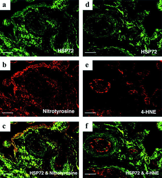
Double-immunofluorescent labeling showing the relative localization of HSP 72 and nitrotyrosine (a–c), and 4-HNE (d–f), respectively, in villous tissues experiencing 20 minutes of hypoxia, followed by 2 hours of reoxygenation with air/5% CO2. Note a mild increase in fluorescent signal of 4-HNE in the syncytiotrophoblast that was not disclosed by the chromogenic technique (e). Furthermore, HSP 72 is a more general marker of oxidative stress than nitrotyrosine and 4-HNE. Scale bar, 50 μm.