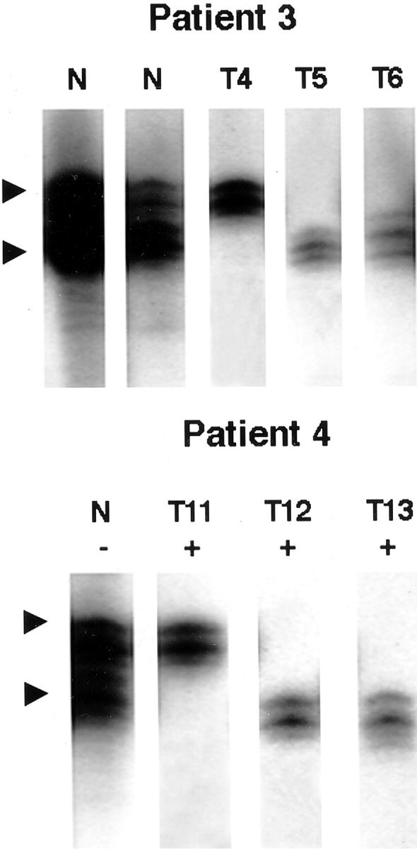
Figure 2.

Representative results of the X-chromosome inactivation analysis of 10 tumors in two female patients. The X-chromosome inactivation method (HUMARA) was used to evaluate clonality of separate tumors. In patient 3, uterine tumor T4 shows methylation of the upper allele and methylation of the lower alleles in uterine tumors T5 and T6. In patient 4, uterine tumor T11 shows methylation of the upper allele, and uterine tumors T12 and T13 show lower allele methylation. At least two of the uterine tumors in each patient arise independently as a different clone. Tumor number corresponds to the tumor number in Table 2▶ . N, normal control, undigested (−) or digested (+), with restriction endonuclease HpaII.
