
Figure 1.
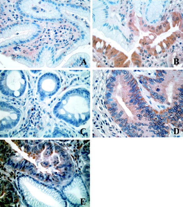
Immunohistochemical analysis of PLC δ2 expression in human gastric healthy (A), type I IM (PLC δ2-positive) (B), type I IM (PLC δ2-negative) (C), type II IM (D), and gastric adenocarcinoma (E). Note in B and E the specificity of the reaction in different glands of the same microscopic field.