
Figure 3.
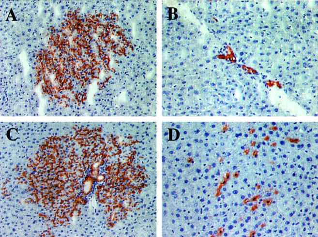
Morphological appearance of proliferated cell clusters at 6 months after transplantation of ED 14 FLEP cells. A: Moderately large hepatocytic cluster. B: Cluster of mature bile duct cells that have proliferated within a host bile duct next to a venous channel. C: Large mixed cluster of transplanted cells containing both hepatocytes and well-differentiated bile ducts. At the periphery of the cluster, cells with a hepatocytic phenotype appear to be extending into the surrounding parenchyma. D: Endothelial cell cluster. Cells with a small nucleus and extensive DPPIV+ cytoplasm are loosely interspersed between hepatocytes and appear to be within the liver sinusoids. By immunohistochemistry, these cells co-stain for rat endothelial cell antigen, RECA-1 (data not shown). Original magnifications: ×100 (A and C); ×200 (B and D).