
Figure 8.
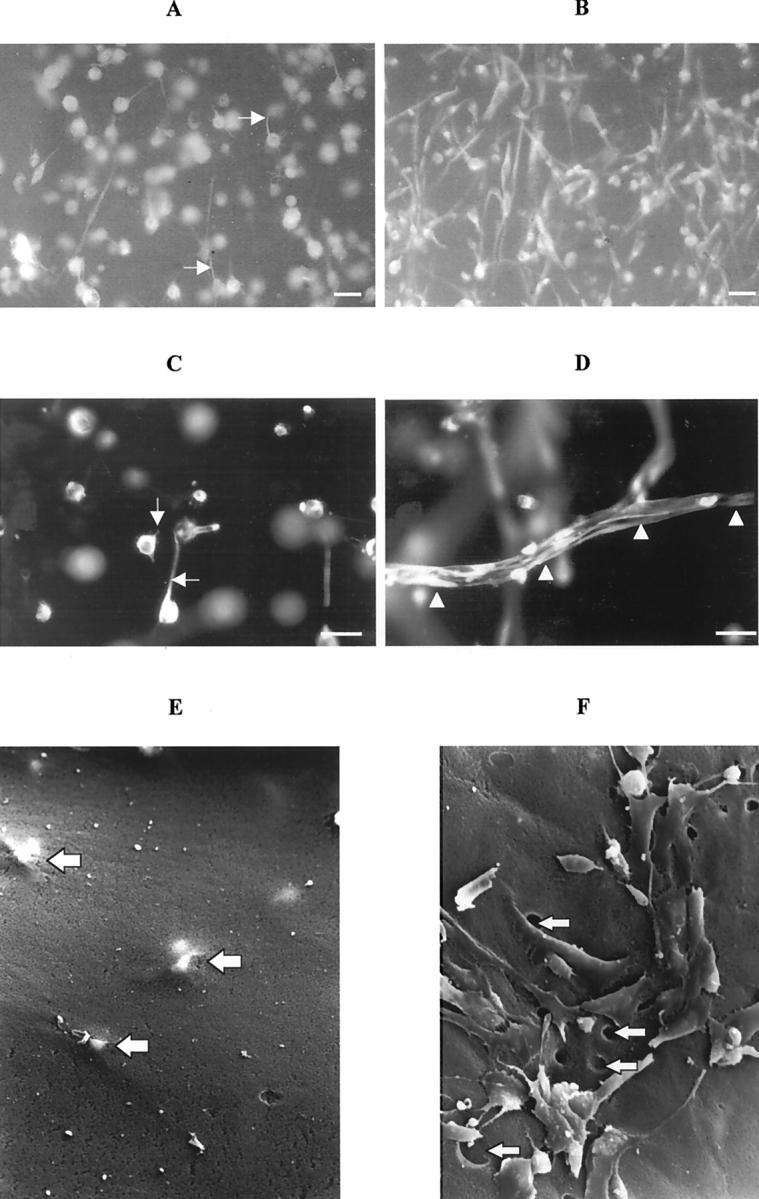
Photomicrographs of whole matrices fluorescently immunostained for CK18 (A–D). During collagen matrix contraction the cells were initially round with small processes extending within the matrix (A; day 1) and were later seen to adopt a stellate and spindle-shaped appearance by day 7 (B). The cells in matrices incubated with Galardin only extended out small processes into the surrounding matrix (C, arrows; day 1) whereas in control cell-populated matrices, the cells characteristically were stellate in appearance and even formed tunnels within the matrix (D, arrowheads; scale bars, 10 μm). Electron micrographs of matrices (E and F) illustrating that no holes or cells are present on the matrix surface, early in the contractile process (day 1; F). RPE cells can still be visualized just beneath the matrix surface (F, large arrows). By the seventh day (E), cells within untreated matrices had reached the matrix surface and numerous empty holes could be seen (E, arrows; original magnification, ×1150).