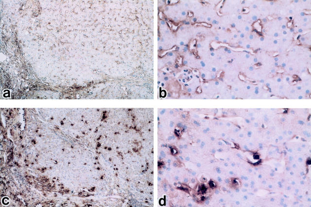
Figure 2.
Light microscopic distributions of endothelin A receptor (ETAR) and endothelin B receptor (ETBR) in human cirrhotic liver using immunoperoxidase staining. a and b: Immunoperoxidase-positive substance for ETAR remains weakly detectable compared to control liver but remains low in level (a: ×100; b: ×400). c and d: Reaction products of ETBR are markedly increased on sinusoidal endothelial cells and hepatic stellate cells compared to control human liver (c: ×100; d: ×400). Hematoxylin counterstain.

