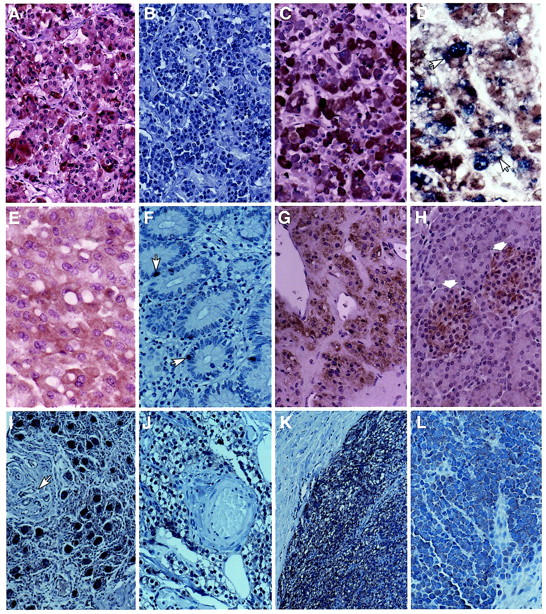
Figure 1.

Immunohistochemical localization of myosin XVA in neuroendocrine tissues. A: Normal anterior pituitary staining strongly (3+) with antibody PB48 in most cells. B: Preabsorption of the antibody with 10 μg/ml purified antigen abolished staining. C: Antibody PB78 also produced strong (3+) staining in normal anterior pituitary cells. D: Combined staining with anti-GH antibody (blue) and myosin XV showed localization of myosin XVA in some GH-producing cell (arrows). E: ACTH adenoma showing diffuse positive staining with PB78. F: Normal endocrine cells (arrows) in the ileum are positive for myosin XV with the PB48 antibody. G: The adrenal medulla shows moderate positive staining (2+) for myosin XVA. H: The islet cells (arrows) are positive (1+) for myosin XVA and the exocrine pancreas is negative. I: The ganglion cells from the retroperitoneum are strongly positive (3+) for myosin XVA. The blood vessel and connective tissues (arrows) are negative. J: Normal parathyroid gland tissue shows positive staining (1+) for myosin XVA. K: Parathyroid adenoma showing strong positive staining (3+) with PB48 while the adjacent fibrous connective tissue is negative. L: Merkel cell carcinoma showing positive cytoplasmic staining (1+) for myosin XVA. Original magnifications: ×250 (A–D, F–L), ×300 (E).
