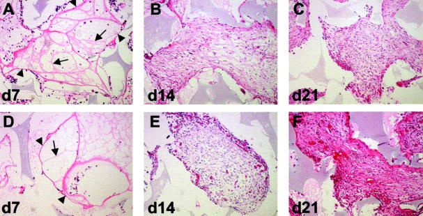
Figure 2.
Increased neovascularization and tissue density in TSP2-null animals. Shown are representative sections of PVA sponge implants recovered at 7 (A, D), 14 (B, E), and 21 (C, F) days from control (A–C) and TSP2-null (D–F) mice and stained with H&E. Fine invasion (d7) characterized by thick (arrowheads) and thin (arrows) fibrovascular bundles was similar in both genotypes (A, D). Dense invasion was characterized by increased angiogenesis and tissue density in TSP2-null animals (E, F). Differences between genotypes were more pronounced at 21 days (compare C and F). Original magnifications, ×100.

