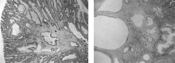
Figure 5.
H&E-stained slides of juvenile polyps. Left: A juvenile polyp (original magnification, ×2.5) from a SMAD4 mutation carrier (family 20). Note areas that look hyperplastic (a) and areas of classical juvenile polyp morphology, with expanded cysts and normal epithelium (b). Right: A classical juvenile polyp (original magnification, ×2.5) from a SMAD4 mutation-negative patient (family MD) with morphology of type (b).

