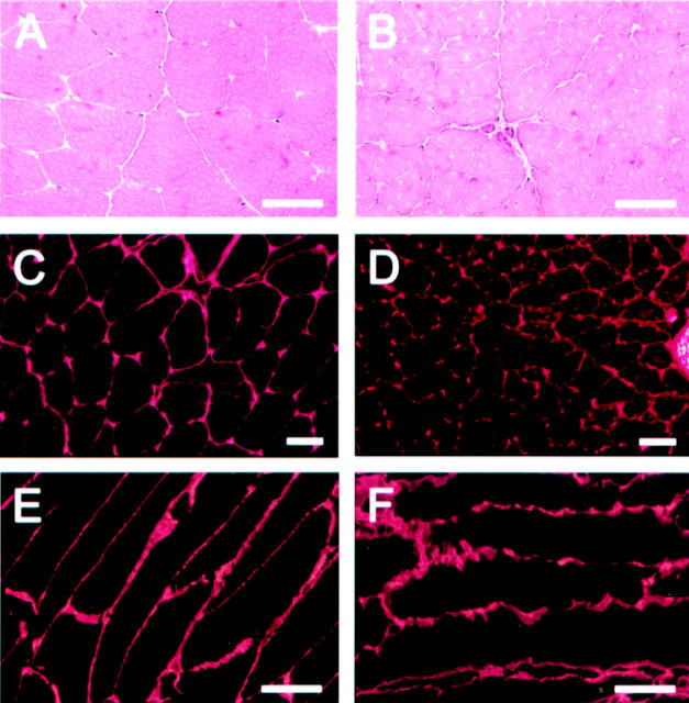
Figure 4.
H&E and immunofluorescence stainings of muscles from wild-type mice (A, C, and E) and Col13a1N/N mice (B, D, and F). The muscle fibers of the mutant mice have an uneven appearance in the H&E stainings, with rough edges (B). Staining with anti-XIII/NC3 antibodies, which detect the ectodomain of type XIII collagen, reveal clear expression of this collagen type in the Col13a1N/N mice (D), but the myofiber structure is uneven compared with that in the wild-type mice (C). Smooth, linear staining of longitudinal muscle sections with anti-type IV collagen antibodies is seen in the wild-type (E), whereas the basement membranes between the muscle fibers are highly uneven and fuzzy in appearance in the mutant case (F). Scale bars, 20 μm.

