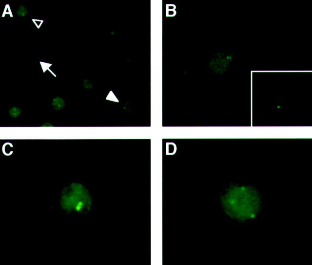
Figure 1.
Representative abnormal centrosome staining in SK-MES-1 with significant CIN. Centrosomes were considered abnormal if there were three or more signals and/or the diameter was more than three times larger than that of human normal fibroblasts. Representative SK-MES-1 cells exhibiting normal centrosome staining (arrow in A, enlarged figure in B) and abnormalities in size (open arrowhead in A, enlarged figure in C) or numbers (solid arrowhead in A, enlarged figure in D). Representative centrosome staining of normal fibroblasts is shown at the same magnification in the inset of B.

