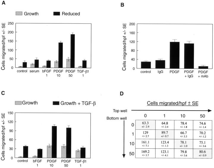
Figure 3.
Transdifferentiated cells exhibit increased migration in response to PDGF-BB. A: Av 17 cells cultured in growth medium (hatched bars) or in reduced medium (solid bars) for 6 days were tested for the ability to migrate toward EBM with 0.1% bovine serum albumin (control), EBM with 5% FBS (serum), 1 ng/ml bFGF, 10 ng/ml PDGF-BB, 50 ng/ml PDGF-BB, or 1 ng/ml TGF-β1. B: A neutralizing anti-PDGF-BB monoclonal antibody (mAb) and an isotype-matched IgG were tested for the ability to block the migration of av 17 cells, which were grown in reduced medium for 6 days, toward 10 ng/ml PDGF-BB. C: Av 15 cells cultured in growth medium (hatched bars) or in growth medium with 1 ng/ml TGF-β1 (solid bars) for 6 days were tested as in A. D: Checkerboard analysis was performed on av 17 cells grown in reduced medium for 6 days. PDGF-BB at 0, 1, 10, or 50 ng/ml was added to top and bottom wells as indicated, and cells allowed to migrate for 4 hours. The shaded boxes on the diagonal highlight the finding that PDGF-BB does not elicit random cell migration toward increasing concentrations of PDGF-BB in the upper and lower chambers.

