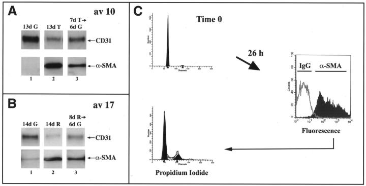
Figure 4.
Characterization of endothelial transdifferentiation. A: Av 10 cells were cultured in growth medium (G) (lane 1) or in growth medium with 1 ng/ml of TGF-β1 (T) for 7 days (lanes 2 and 3). After 7 days, cells were trypsinized and replated on new gelatin-coated dishes in growth medium (lanes 1 and 3) or in growth medium with TGF-β1 (lane 2) for 6 additional days. B: A similar time series was used with av 17 cells and reduced medium (R) instead of TGF-β. Cell lysates were analyzed by Western blot using anti-CD31 and anti-α-SMA. C: Av 17 cells were grown in reduced medium for 6 days. A portion of the cells was stained with propidium iodide to determine the cell-cycle distribution (top left). The rest of the cells were replated in growth medium containing 10% FBS and 2 ng/ml of bFGF at 30,000 cells/cm 2 for 26 hours. The cells were then harvested, permeabilized with 0.1% Triton-X-100 for immunostaining of intracellular α-SMA by flow cytometry (right), and simultaneously stained with propidium iodide (bottom left). The gray line at the right represents cells stained with an isotype-matched IgG2a control.

