
Figure 1.
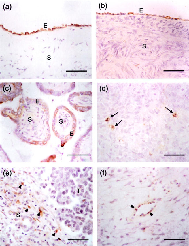
Immunohistochemical analysis and localization of SPARC expression in normal ovary and ovarian tumors. Photomicrographs are taken from sections of normal ovary (a), serous benign ovarian adenoma (b), borderline ovarian tumor (c), and grade III serous ovarian carcinoma (d, e, and f). All sections were stained with a rabbit polyclonal antibody specific to SPARC. Immunoreactive cells, which are stained brown in the cytoplasm, are found in the epithelium (E) of normal ovary, benign ovarian adenoma, and borderline ovarian tumor (a, b, and c). No staining is observed in the underlying stroma (S) of these tissue samples. In grade III serous ovarian carcinoma (d), immunoreactivity is significantly reduced in which only a few scattered cancer cells are stained brown (arrows). e: In contrast to normal ovary, benign and borderline ovarian tumors, scattered positively stained cells (arrowheads) can be found in the stroma (S) of ovarian cancer adjacent to the tumor (T). Endothelial cells in ovarian carcinoma also show occasional positivity to SPARC staining (arrowheads; f). Scale bars, 50 μm.