
Figure 2.
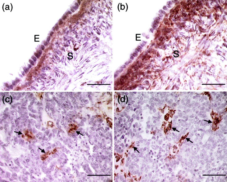
Localization of SPARC immunoreactivity in normal ovary (a and b) and ovarian cancer (c and d) using the monoclonal antibody AON-5031. a: Very weak staining was observed in the normal ovarian epithelium (E). In contrast, cells in the underlying stroma (S) showed strong SPARC immunoreactivity. b: The staining of stromal cells was intensified after heating with antigen-unmasking solution. However, the normal ovarian epithelial cells remain faintly stained. c: Positive staining was seen in a few cancer cells and some scattered stromal cells (arrows). No immunoreactivity was evident in the vast majority of cancer cells. d: Stromal cells (arrows) showed strong SPARC staining after antigen retrieval, whereas cancer cells remain unstained. Scale bars, 50 μm (a and b); 100 μm (c and d).