
Figure 7.
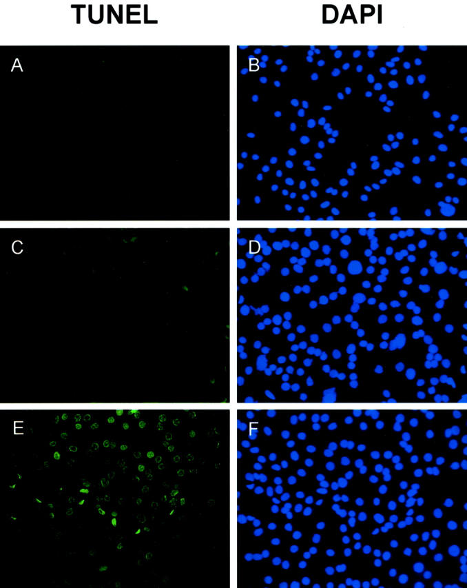
Detection of apoptotic ovarian cancer cells by TUNEL assay. SKOV3 cells seeded in chamber slides were treated with different concentrations of human blood platelet SPARC proteins for 48 hours (A and B, untreated; C and D, 1 μg/ml; E and F, 5 μg/ml). After incubation, TUNEL assays were performed (A, C, and E). To visualize the cell nuclei regardless of apoptosis, SKOV3 cells were also stained by DAPI (0.5 μg/ml) at room temperature for 10 minutes (B, D, and F). Early apoptotic cells and DAPI-stained cells were detected by fluorescence microscopy. DAPI-stained cell nuclei were seen as blue dots whereas the nuclei of apoptotic cells were stained green. There is a significant increase in the number and staining intensity of apoptotic cell nuclei when SKOV3 cells were treated with increasing amounts of SPARC, indicating more DNA strand breaks.