Figure 2.

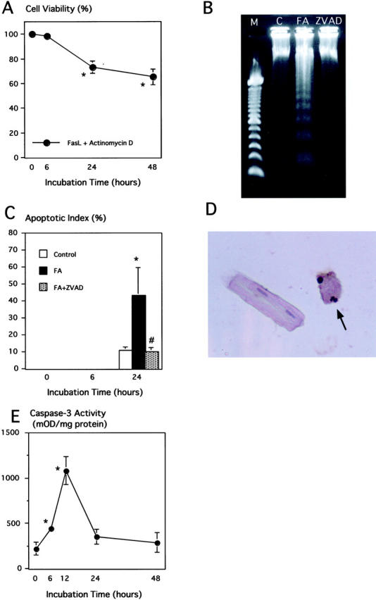
Effect of Fas stimulation on viability, DNA fragmentation, and caspase-3 actiivty of cultured adult cardiomyocytes. A: The percentage of the dye-excluding cells were calculated in proportion to the vehicle control at the corresponding time. *, P < 0.05 compared with the control. B: Gel electrophoresis of DNA extracted from cultured cardiomyocytes incubated for 24 hours with normal buffer (C), FasL plus actinomycin D (FA), or FA plus zVAD.fmk (ZVAD). Lane M indicates 100-bp marker ladder. Lane FA shows a clear ladder pattern. C: Apoptotic index of Fas-stimulated cardiomyocytes at 0, 6, and 24 hours after incubation. Note that there was no evidence of TUNEL-positive cells at 0 and 6 hours. FA, FasL plus actinomycin D; ZVAD, FA plus zVAD.fmk. *, P < 005 versus control; #, P < 0.05 versus FA. D: A TUNEL-positive (closed arrow) and negative adult cardiomyocytes (open arrows) after 24 hours incubation with FasL plus actinomycin D. Original magnification, ×400. E: Quantitative analysis of caspase-3 activity in cultured adult cardiomyocytes treated with FasL plus actinomycin D. Caspase-3 activity was measured using DEVD-pNa; n = 5. *, P < 0.05 compared with control.
