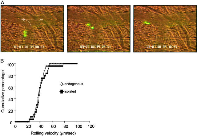
Figure 5.
A: Rolling of isolated neutrophils in vivo. Sequential video frames (left to right) show isolated neutrophils rolling along the vascular endothelium. Cells were fluorescently labeled with CFDA-SE and intravital microscopy performed on the murine cremaster microcirculation ∼30 minutes after surgery. QuickTime movies of these rolling cells are available on the website. B: Rolling velocities of isolated neutrophils and endogenous leukocytes in vivo. Images of rolling cells were recorded onto videotape immediately after injection of isolated neutrophils, ∼30 minutes after surgical preparation of the recipient mice. Velocities were determined by measuring distance traveled over successive video frames for a period of at least 2 seconds per cell. Data are presented as cumulative velocity histograms for at least 20 cells per histogram.

