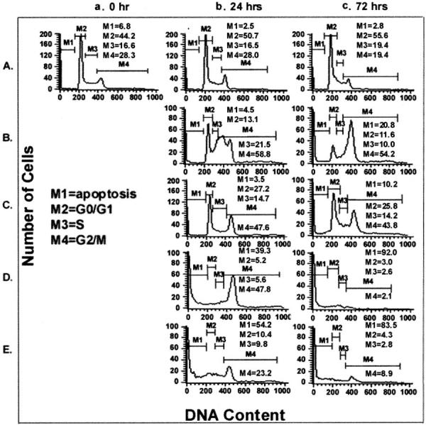
Figure 3.

Cell-cycle analysis of CEM-6 cells after various treatments. CEM-6 cells were exposed to various agents as shown in Figure 2 ▶ [untreated controls (A), cisplatin (B), VP-16 (C), vincristine (D), and irradiation (E)]. Cells were collected for analysis at 0 (a), 24 (b), and 72 (c) hours after treatment.
